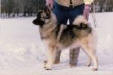
According to the OFA (Othopedic Foundation for Animals) ... of 3384 Norwegian Elkhounds xrayed from 1974 to 2006, 6.8% of the hips were excellent and 19.6% were dysplastic.
Our breed is ranked 33rd for this disease.
Hip Dysplasia is a genetic disease because of the various degrees of arthritis (also called degenerative joint disease, arthrosis, osteoarthrosis) it can eventually produce, leading to pain and debilitation.
The very first step in the development of arthritis is articular cartilage (the type of cartilage lining the joint) damage due to the inherited bad biomechanics of an abnormally developed hip joint. Traumatic articular fracture through the joint surface is another way cartilage is damaged. With cartilage damage, lots of degradative enzymes are released into the joint. These enzymes degrade and decrease the synthesis of important constituent molecules that form hyaline cartilage called proteoglycans. This causes the cartilage to lose its thickness and elasticity, which are important in absorbing mechanical loads placed across the joint during movement. Eventually, more debris and enzymes spill into the joint fluid and destroy molecules called glycosaminoglycan and hyaluronate which are important precursors that form the cartilage proteoglycans. The joint's lubrication and ability to block inflammatory cells are lost and the debris-tainted joint fluid loses its ability to properly nourish the cartilage through impairment of nutrient-waste exchange across the joint cartilage cells. The damage then spreads to the synovial membrane lining the joint capsule and more degradative enzymes and inflammatory cells stream into the joint. Full thickness loss of cartilage allows the synovial fluid to contact nerve endings in the subchondral bone, resulting in pain. In an attempt to stabilize the joint to decrease the pain, the animal's body produces new bone at the edges of the joint surface, joint capsule, ligament and muscle attachments (bone spurs). The joint capsule also eventually thickens and the joint's range of motion decreases.
No one can predict when or even if a dysplastic dog will start showing clinical signs of lameness due to pain. There are multiple environmental factors such as caloric intake, level of exercise, and weather that can affect the severity of clinical signs and phenotypic expression (radiographic changes). There is no rhyme or reason to the severity of radiographic changes correlated with the clinical findings. There are a number of dysplastic dogs with severe arthritis that run, jump, and play as if nothing is wrong and some dogs with barely any arthritic radiographic changes that are severely lame.
Many Norwegian Elkhounds also are quite prone to Sebaceous Cysts:
Sebaceous, or oil-producing, glands sometimes become plugged with gland material and other debris, which can lead to a bacterial infection; it is similar to the development of acne. Sebaceous cysts are not terribly serious, although if large enough they may cause pain from the pressure. Sometimes they can be treated conservatively by opening the cyst and treating the secondary infection. Warm soaks or hot packs followed by firm pressure may allow the cyst to open. The material inside can then be squeezed out daily. If, at the same time, your dog could be put on an inexpensive oral antibiotic, you may be able to avoid the need for surgery.
Large cysts that refill repeatedly can be successfully removed. This is especially important for outdoor dogs, whose draining cysts may attract flies. If you opt for removal, your veterinarian can lance the cyst using only a local anesthetic or can do a more extensive excision using general anesthesia.
Author(s): Wilcox, Bonnie, D.V.M.
Publication: Dog Fancy
Issue Date: June 1993